What’s the Difference Between a Magnifying Glass and a Dermatoscope?

In this post, we’ll break down what each tool is, what it can realistically show, and when it’s worth moving from a simple magnifying glass close-up to a dermatoscope-based assessment.
What is a Dermatoscope?
A dermatoscope is a handheld clinical tool used to examine moles, lesions, and fine skin structures up close. Unlike a basic magnifier, it combines magnification with controlled illumination—often with polarized options—to reduce surface glare, so pigment detail and vascular (red) features are clearer and easier to interpret.

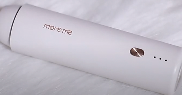
In practice, the real advantage isn’t just “seeing bigger,” but creating repeatable, comparable images—especially when you’re documenting a baseline and checking changes over time. For example, devices like the iMoreMe Dermascope tools are designed for consistent documentation, offering a 2MP, 1920×1080 medical-grade camera, up to 50× macro magnification, and four-spectrum viewing (daylight, parallel polarization, cross polarization, and UV) to capture different details under controlled conditions. Features like WiFi connectivity, AI-generated reports, and cloud backup can also simplify workflow—making it easier to save cases, compare follow-ups, and share findings clearly with clients or within a team.
What is a Magnifying Glass?
A magnifying glass is a simple handheld lens that makes objects look bigger by bending light—basic optical magnification. Because it doesn’t include its own lighting, what you see can change a lot depending on the room light, your angle, and the lens quality. With bright, even lighting, it can be surprisingly useful; with harsh overhead light or shadows, details can be easy to misread.

For skincare or everyday checks, it’s best for surface-level observations—dry flakes, leftover makeup, visible pore openings, rough patches, or obvious blackheads. But it’s still limited: it mainly enlarges what’s on the surface and can be affected by glare, so it won’t consistently reveal the deeper pigment or vascular patterns that clinicians rely on when doing more advanced skin assessments.
Dermatoscope vs. Magnifying Glass Core Differences at a Glance
|
Feature |
Magnifying Glass |
Dermatoscope |
|
Purpose |
General viewing of surface details |
Clinical assessment of skin lesions and structures |
|
Magnification & clarity |
Simple lens; clarity varies by lens quality and angle |
Optimized optics designed for skin examination; clearer detail |
|
Lighting |
Relies on ambient room light |
Built-in, controlled illumination (often polarized) |
|
Surface vs. subsurface detail |
Mostly surface-level; limited structural visibility |
Enhanced visibility of deeper pigment/vascular patterns and structures |
|
Contact method |
Typically non-contact |
Can be contact or non-contact (depends on device/mode) |
|
Image capture |
Optional phone clip; not standardized |
Often supports digital documentation (device-dependent) |
Dermatoscopes and Magnifying Glass Use Cases
1.When a dermatoscope is the better choice
A dermatoscope is the better option when surface detail isn’t enough—especially for moles or lesions where subtle patterns matter. Because it uses controlled illumination (often polarized), it reduces glare and makes underlying structures easier to read, including pigment patterns and vascular (red) features that a simple lens can’t show reliably.

It’s also the stronger choice when you want consistency over time. Many dermatoscopes can capture more standardized images, so follow-ups are easier to compare—you're looking at the same area under similar conditions, rather than relying on memory or slightly different lighting and angles. That’s what makes it valuable in higher-stakes situations where small changes are worth tracking carefully.
2.When a magnifying glass is “good enough”
For everyday skincare checks—at home or in a basic consult—a magnifying glass is usually enough to confirm what’s happening on the surface. With bright, even lighting, it helps you see fine flaking, rough patches, visible pore openings, leftover product, or obvious blackheads more clearly. It’s best thought of as a “closer look,” not a deeper diagnostic tool.

It’s also useful for low-stakes progress checks. If you’re mainly trying to answer questions like “Is my dryness improving?” “Does this area look calmer?” or “Is texture smoothing out?” a magnifying glass can give you quick, practical feedback—no special lighting or formal photo setup required.
Limitations & Common Misunderstandings When Using a Magnifying Glass or Dermatoscope
1.Magnification ≠ diagnosis
Seeing more detail doesn’t automatically explain what you’re looking at. A magnifying glass can make surface changes easier to notice—flaking, roughness, visible pore openings, leftover product—but it doesn’t reliably tell you what’s driving them. A lot of issues can mimic each other up close (dryness vs. irritation, early inflammation vs. simple sensitivity, clogged pores vs. tiny milia), and basic magnification can’t reveal the deeper structural cues that help you sort those apart with confidence.

It can also make normal skin look “worse” than it is. Under close-up viewing, pores, peach fuzz, and fine lines can seem more dramatic—especially under harsh lighting or at a sharp angle. That’s why it helps to keep perspective: check under consistent lighting, step back to see how it looks at normal distance, and factor in what the person feels (tightness, stinging, itch, sensitivity). Otherwise, you risk treating a normal feature as a problem—and creating irritation in the process.
2.A dermatoscope is powerful, but context still matters
A dermatoscope tool can reveal much richer detail than a simple lens, but it still isn’t an “instant answer.” The patterns it shows need trained interpretation, and clinicians rarely judge them in isolation—they pair what they see with the story behind it: how long it’s been there, whether it itches or bleeds, and whether it’s changing in size, shape, or color over time.
Used casually, dermoscopy can push people toward extremes: false reassurance (“it looks okay up close”) or unnecessary alarm (“I see a pattern, so it must be serious”). A safer approach is to treat a dermatoscope tool as a tool for clearer viewing and documentation—not a substitute for medical evaluation when a spot is new, changing, asymmetric, bleeding, painful, or otherwise unusual.
Read more:
https://www.djmimoreme.com/resources/blog/can-a-dermoscopy-detect-cancer.html
https://www.djmimoreme.com/resources/blog/how-accurate-are-dermatoscopes.html
Conclusion
A magnifying glass is great for quick, surface-level checks—useful, accessible, and often all you need for everyday skincare observations. A dermatoscope, on the other hand, is designed for clinical-level viewing and consistent documentation, making it the better option when patterns beneath the surface matter and when follow-up comparisons are important.
Table of Contents
Related information


How can we help you?
Have specific questions or requests? Fill out our inquiry form, and our dedicated team will get back to you promptly. Your inquiries are important to us, and we are committed to providing comprehensive and personalized responses tailored to your needs.


 Reach out to us today!Whether you are preparing to start your own
business in the beauty industry, are ready to upgrade your
equipment, or are just interested in our products,Contact us
today, and let’s explore how we can partner to achieve your goals
and drive your success to new heights!
Reach out to us today!Whether you are preparing to start your own
business in the beauty industry, are ready to upgrade your
equipment, or are just interested in our products,Contact us
today, and let’s explore how we can partner to achieve your goals
and drive your success to new heights!