What is a dermatoscope, and what does it see?

A dermatoscope isn't just a magnifying tool—it's a window into the hidden architecture of our skin. This article explores what a dermatoscope actually sees, how it works, and why it's reshaping the standard of care in skin diagnostics.
Learn more:
https://www.djmimoreme.com/what-is-a-dermascope-used-for
1.What Is a Dermatoscope?
A dermatoscope is a specialized handheld device designed to provide magnified, illuminated visualization of the skin’s microstructures, particularly at the epidermal and dermo-epidermal junction. Unlike a standard magnifying lens, a dermatoscope combines high-resolution optics with polarized or non-polarized lighting to eliminate surface reflection and enhance the visibility of subsurface pigmentation, vascular networks, and structural asymmetries.
Rather than relying on the naked eye, which is limited to surface-level observation, dermoscopy allows clinicians to examine pigmentation depth, irregular melanocytic patterns, and early morphological changes that may indicate malignant transformation. This level of detail is critical when evaluating suspicious lesions, especially for conditions like melanoma, where early diagnosis significantly improves patient outcomes.
Modern dermatoscopes often integrate with digital imaging systems, enabling real-time capture, storage, and comparison of skin lesions over time—transforming skin checks into a data-driven diagnostic process.
Learn more:
https://www.djmimoreme.com/what_is_dermoscopy_and_why_is_it_important
2.What Does a Dermatoscope Actually See?
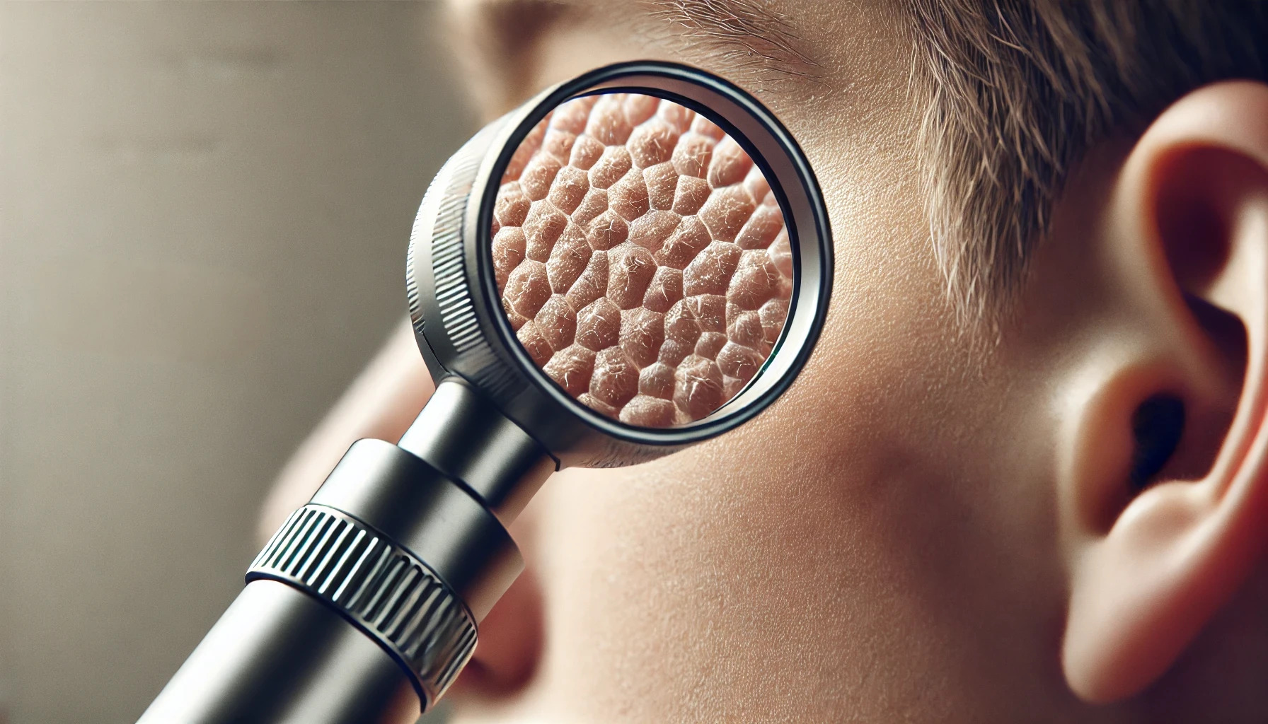
A dermatoscope opens a visual gateway beneath the skin’s surface—beyond what the human eye can perceive—revealing structural clues that help differentiate benign from malignant lesions. In the context of melanoma detection, dermoscopy tool highlights subtle patterns and chromatic shifts associated with atypical melanocytic activity.
Rather than focusing on surface appearance, the device uncovers features located from the stratum corneum down to the dermal papillary layer, where early signs of melanoma often begin.
The table below summarizes key dermoscopic signs often associated with early melanoma detection:
|
Feature |
What It Suggests |
|
Atypical pigment network |
Irregular pigmentation—possible melanoma |
|
Irregular dots/globules |
Uneven shapes and distribution—warning sign |
|
Blue-white veil |
Deep pigmentation—linked to invasive growth |
|
Asymmetric streaks |
Uneven radial lines—may indicate melanoma |
|
Regression structures |
White/gray areas—possible immune response |
These visual features, when interpreted in combination and context, can dramatically improve diagnostic accuracy. Dermoscopes don’t replace histopathology, but they provides a critical first layer of decision-making—especially when applied with experience and pattern recognition.
3.Clinical Advantages: Dermatoscope vs the Naked Eye
3.1 Improved Early Detection Accuracy
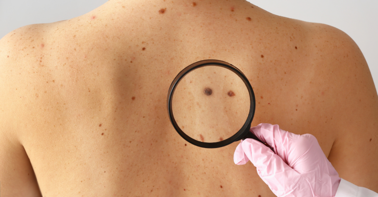
Standard visual skin checks rely on surface-level observation and subjective judgment. While trained eyes can identify some suspicious lesions, many early melanomas appear deceptively benign to the naked eye. Dermatoscopes, however, provides magnified, light-enhanced visualization of subsurface structures—allowing clinicians to detect subtle irregularities such as atypical pigment networks, blue-white veils, and vascular patterns that would otherwise go unnoticed.
This enhanced visibility translates into significantly higher diagnostic sensitivity, especially for early-stage melanomas, which are most treatable when caught early.
3.2 Fewer Unnecessary Biopsies
While over-diagnosis is a common concern in skin cancer screening, dermoscopy tool can help strike the right balance between caution and clinical accuracy. By offering a clearer visual distinction between benign and malignant features, it reduces the likelihood of overreacting to harmless moles or freckles.
This means fewer patients are subjected to unnecessary biopsies, which not only reduces physical scarring but also lowers healthcare costs and minimizes psychological stress associated with false alarms.
3.3 Better Surgical Planning
When a lesion is confirmed or strongly suspected to be malignant, precise excision is critical. Dermoscopy tool aids in mapping the true clinical margins of a lesion—especially in cases where the surface appearance doesn’t reveal the full extent of subclinical spread.
This ensures more complete removal of cancerous tissue, minimizing the chance of recurrence while also preserving as much healthy skin as possible—a key concern for facial lesions or cosmetically sensitive areas.
3.4 Effective Long-Term Monitoring
Not all atypical lesions require immediate removal. For patients with multiple moles or a history of melanoma, digital dermatoscopes enable long-term tracking of skin changes. High-resolution images captured over time can be compared side-by-side to monitor size, color, or structural evolution.
This serial imaging approach is especially valuable for borderline lesions that don’t meet current surgical criteria but may evolve into malignancy later. It also provides objective documentation for follow-ups.
Clinics in Germany and Italy are leading the way by integrating AI-supported dermoscopic platforms that flag suspicious changes over time. These systems assist dermatologists in recognizing early warning signs, even before visible symptoms arise.
3.5 Enables Teledermatology and Remote Access
Dermatoscope is also revolutionizing access to quality dermatologic care in underserved areas. When paired with digital cameras or smartphone attachments, dermoscopic images can be securely transmitted to specialists anywhere in the world—making teledermatology a practical solution for early triage.
Patients in rural or low-resource settings no longer have to wait weeks or travel long distances to see a dermatologist. Remote experts can assess dermoscopic images and recommend timely next steps.
3.6 Bridges the Gap Between Subjectivity and Data
One of the most transformative aspects of this tool is its ability to convert what was once a subjective assessment into a data-driven, pattern-based diagnostic process. By providing standardized visual criteria, dermoscopy empowers clinicians to base decisions on reproducible structures rather than intuition alone.
When combined with digital recordkeeping and AI algorithms, dermoscopes create a highly reliable system that improves consistency across practitioners, builds patient trust, and elevates the overall quality of care.
Learn more:
https://www.djmimoreme.com/visia-vs-imoreme-why-imoreme-is-the-best-value-skin-analysis-machine
Conclusion:
As digital integration and AI continue to advance, the dermatoscope is poised not just as a diagnostic tool, but as a cornerstone of precision skin health. Whether in a high-tech clinic or a remote setting via telemedicine, its impact is clear: we can now see what truly lies beneath the surface.
Table of Contents
Related information
How can we help you?
Have specific questions or requests? Fill out our inquiry form, and our dedicated team will get back to you promptly. Your inquiries are important to us, and we are committed to providing comprehensive and personalized responses tailored to your needs.


 Reach out to us today!Whether you are preparing to start your own
business in the beauty industry, are ready to upgrade your
equipment, or are just interested in our products,Contact us
today, and let’s explore how we can partner to achieve your goals
and drive your success to new heights!
Reach out to us today!Whether you are preparing to start your own
business in the beauty industry, are ready to upgrade your
equipment, or are just interested in our products,Contact us
today, and let’s explore how we can partner to achieve your goals
and drive your success to new heights!